Amyloidosis
Slide #10
This 91-year-old female had a history of hypertension, type II diabetes mellitus, congestive heart failure and atrial fibrillation. She was found unresponsive by the staff of the nursing home in which she resided, and was brought toCPMCbyEMS. Resuscitation attempts were unsuccessful.
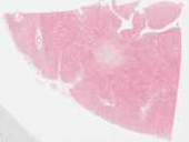
The myocardium in this section is widely infiltrated by an amorphous, pink extracellular material (amyloid). Broad fields and aggregates of amyloid separate myocytes, in some instances extremely widely. In contrast to fibrous tissue (scar) note that there are no cell nuclei within the amorphous protein aggregates, and the amyloid is not well resolved microscopically into fibrils or filaments (this would be seen on transmission electron microscopy).
Checklist: Have you identified
- amyloid in the interstitial spaces
- myocytes
Questions
- Define "amyloidosis"
- What are the main types of amyloidosis?
- How does the pathologist document that the extracellular material is amyloid?
- What type of amyloid was probably present in this patient?