Acute Endocarditis
Slide #11
This 64-year-old man had diabetes for 10 years, poorly controlled with oral hypoglycemics. He had developed multiple, non-healing ulcers on his foot and was admitted for debridement of these, but developed septic shock. Amputation of the left lower leg was required, and one week later he developed myocardial infarction, cerebellar haemorrhage with multiple infarcts and left lateral ventricular subarachnoid haemorrhage, which directly caused his death. At autopsy there was acute endocarditis of the mitral valve and a valvular ring abscess, though to have directly contributed to septic emboli to the coronary artery, central nervous system and (at autopsy) to the kidney (with a recent infarct on gross exam).
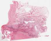
This slide shows the mitral valve with vegetations composed of baterial colonies, thrombotic material and neutrophils. Note that the inflammation also extends through the valve substance deeply into the underlying myocardium. On high power, the bacterial colonies stain blue and individual cocci can be delineated (without using the oil lens!). If your slide contains a longitudinal section through the valve, note the fibrosa and spongiosa layers if possible.
Checklist: Have you identified
- bacterial colonies
- thrombotic material (fibrin, platelets and red blood cells)
- underlying valve tissue (fibrosa, spongiosa layers)
- extension of the infection into the myocardium
Questions
- What were this patient's risk factors for acute endocarditis?
- What were the most likely results of blood cultures?
- Is mitral valve involvement common in infective endocarditis? What are the most commonly involved valves?