Liver I Hepatic Infarct
Slide #3471
C.C.: Elevated serum liver tests 7 days after liver transplantion.
H.P.I.: 50-year-old man with a history of chronic hepatitis C underwent a living-related donor liver transplantation. On post-op day #4 hisLFT's were returning toward normal: T. Protein-5.3 (nl=6.7-8.6 g/dl), alb=3.2 (nl=4.1-5.3 g/dl), t.bili=5.4 (nl=0.3-1.30 mg/dl), d. bili=2.3 (nl=0.04-0.38 mg/dl),AST=70 (nl=12-38 U/L),ALT=202 (nl=7-41 U/L), Alk. phos.=55 (nl=33-96 U/L).
OnPOD#7 hisLFT's were found to be worsening and his immunosuppressants were adjusted in response to acute rejection, diagnosed on liver biopsy.
OnPOD#11 hisLFT's showed T. Protein=4.2, alb=3.1, t. bili=37.7, d.bili=17.0,AST=572,ALT=1750, alk.phos.=311. Ammonia was 49 (nl=11-35 µM/L). Coags remained within normal limits. DiagnosticUSGof the abdomen was performed but could not demonstrate the hepatic artery, despite repeated attempts. Hepatic artery thrombosis with liver infarct was presumed and the patient subsequently underwent a repeat, cadaveric liver transplantation.
Questions
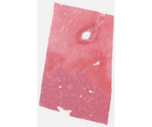
- There are striking regional differences in the staining and other changes in this slide of liver. Describe them.
- In the large area where hepatocytes are no longer visible, what pathologic process has occurred?
- In light of the clinical history, particularly the evidence of hepatic artery thrombosis, describe the sequence of events that led to the pathologic changes in the liver
- In the region of preserved liver parenchyma, what pathologic changes have occurred and what region(s) of the liver is (are) involved (i.e., centrilobular, midzonal, periportal)?
- Which basic type of cell injury is demonstrated in this case?