Acute Myocardial Infarction
Slide #7a
This 62-year-old man underwent heart transplantation for endstage ischemic heart disease. The explant heart grossly showed severe multi-vessel coronary artery disease with grey white patches of scar tissue throughout the ventricular septum and anterior and lateral walls of the left ventricle. Multiple foci of pale myocardium were noted in the anterior wall, which were sampled for microscopy.
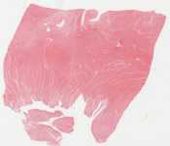
This section shows myocardium with broad areas of fibrosis (due to prior ischemia and infarction). The acutely infracted tissue is recognised on low power by several features, which are each related to the age of the infarct: hypereosinophilic myocytes, myocytes without nuclei and/or without cross-striations, myocytes with contraction bands (due to reperfusion injury) and early neutrophil infiltrates near myocytes with the preceding changes.
Checklist: Have you identified
- Necrotic myocytes
- Neutrophils
- Contraction bands
- Fibrous scars
Questions
Outline the time course and histologic changes in myocardial infarction from the first hours to 6 weeks later.