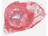
Acute Appendicitis
Slide #3470
C.C.:RLQpain.
H.P.I.: A 68-year-old man with a history ofHTN, NIDDMpresented withRLQpain x 5 hours, increasing in severity, with fevers to 102 F. He reported one episode of non-bilious, non-bloody vomiting on the morning of admission, with continued nausea. He reported having a normal BM on the same morning, and denied a history of diarrhea, hematochezia or melena. His physical exam was notable for T 101.5, P 95, BP 140/85, R 14, a non-distended abdomen with guarding and rebound in theRLQ. Labs demonstrated an elevatedWBCat 17 (nl=3.5-9.0 × 10⁹) with a left shift,PMNs=86%, lymphs=9% (nl:PMN 40-70%, lymphs 20-50). All other lab work was within normal limits. He was taken to theOR,where an appendectomy was performed. He had a satisfactory post-operative course and was discharged home 4 days later.
Questions
- The changes in the appendix involve which portion or portions of the wall?
- Examine the serosa. Describe the major pathologic findings:
- What is the predominant inflammatory cell in the appendix in this case?
- What is the pathogenesis of this appendiceal condition?
- This condition is an example of which of the following: inflammation, repair or neoplasia?
- Should this process be considered acute or chronic, and why?